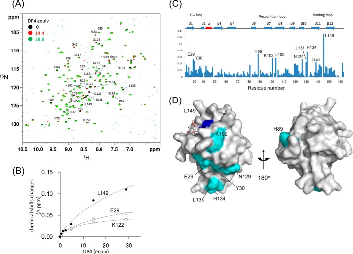
FIGURE 9.
ZG16p binds heparin using its large surface area. A, 1H-15N HSQC spectra of 15N-labeled ZG16p in the absence and presence of heparin tetrasaccharide. B, titration curves for the selected peaks in 1H-15N HSQC spectra of 15N-labeled ZG16p upon addition of heparin tetrasaccharide. C, chemical shift changes of amide signals upon binding to heparin tetrasaccharide. D, mapping of perturbed residues on the ZG16p structure. The residues showing chemical shift change are colored in blue (Δδ > 0.08 ppm) and sky blue (0.04 < Δδ < 0.08 ppm).

