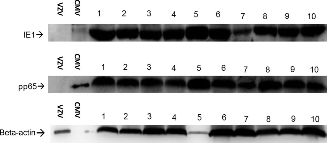
Figure 1. Detection of CMV proteins in primary GBM.
Single cell digests were prepared from de-identified newly diagnosed GBM specimens, washed with PBS and lysates prepared in the presence of proteinase inhibitors. Western blot (WB) analysis was performed using CMV IE1-specific and pp65-specific mAbs on GBM lysates (20 µg per lane). CMV- and VSV-infected fibroblast lysates were used as positive and negative control samples respectively (1 µg per lane). Figure depicts 10 GBM specimens positive for CMV proteins IE1 and pp65. Normal brain lysates obtained from autopsy specimens were negative for detection of CMV proteins (0 out of 5, data not shown).

