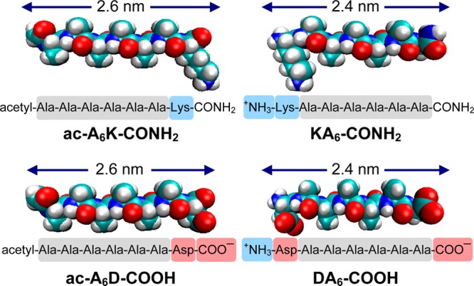
Figure 1.

Molecular modeling, amino acid sequence, and charge distribution of lipid-like self-assembling peptides. The peptide length is similar to biological phospholipids. The hydrophobic domain of the peptides consists of six alanines. Color code: cyan, carbon; red, oxygen; blue, nitrogen; white, hydrogen. Illustrations were generated by VMD. Highlighted domains represent amino acids with positive charge (blue), negative charge (red), and hydrophobic side chains (grey). Using pKa values from the literature the net charge of the peptides at pH 7.4 was calculated to be ac-A6K-CONH2 (+1), KA6-CONH2 (+2), ac-A6D-COOH (−2), DA6-COOH (−1).
