Figure 6.
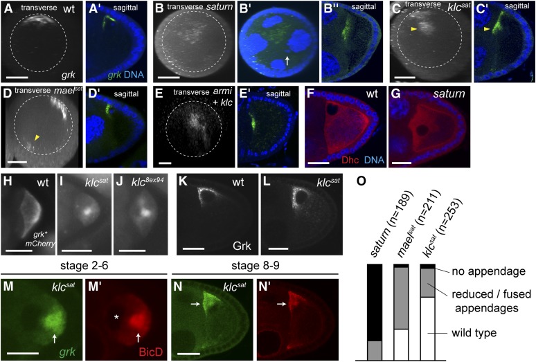
Anterior-central grk mRNA localization in saturn is due to joint inactivation of klc and piRNA pathway. (A−E) Sagittal views as well as transverse projections of a series of z-stack confocal images of endogenous grk mRNA visualized by in situ hybridization in wildtype (A), saturn (B), klcsat (C), maelsat (D), and armi3R13 klc8ex94 double mutant (E) stage 8−10 egg-chambers. Nuclear DNA in the saturn oocyte is marked by arrow (B′), showing that the majority of grk mRNA is not associated to the nucleus. Approximately one-half the grk mRNA (arrowhead, C) is internalized in klcsat germline clones. grk mRNA is also seen in the opposite side of the anterior periphery (arrowhead, D) in maelsat germline clones. (F, G) Anti-Dhc staining in wild-type (F) and in saturn (G) stage 8−9 egg chambers. Dhc is excluded from the nucleus, thus showing the mispositioned nucleus. (H, I, J) grk*mCherry localization in stage 6 oocytes. (H) wild-type (wt), (I) klcsat, and (J) klc8ex94 showing internally localized grk mRNA in klc mutants. (K, L) Anti-Grk staining in stage 8−9 egg-chambers. (K) wildtype (wt). (L) klcsat, showing that Grk translation is not disrupted in klc mutant. (M, N) Localization (arrows) of grk*mCherry (grk, M and N) and the dynein cofactor, BicD (M′ and N′) indicates that dynein-dependent grk mRNA transport is not affected in klcsat germline clone. BicD and grk mRNA are excluded from the oocyte nucleus, showing a posteriorly mislocalized nucleus before its migration to the dorsoanterior corner. (O) Frequencies of abnormal or absent dorsal appendages in mature eggs of indicated genotypes. Scale bars = 5 μm in (M) and 20 μm in the rest.

