
Figure 1. DNA substrates and purified Msh2-Msh3 and Msh2-msh3Y942A.
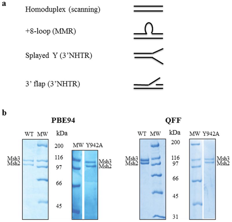
(a) The four different synthetic DNA substrates used in this study were homoduplex (non-specific), +8-loop (MMR), splayed Y and 3′flap (3′ NHTR) substrates. (b) Purified Msh2-Msh3 and Msh2-msh3Y942A (1.5 μg complex each) using the PBE94 purification (left) or the Q-Sepharose Fast Flow purification protocol (right). The protein complexes were analyzed by SDS-PAGE (8%) and stained with Coomassie Blue. Msh2 and Msh3 are indicated. The sizes of molecular weight markers (MW; Bio-Rad, broad range) are indicated alongside the gels in kDa.