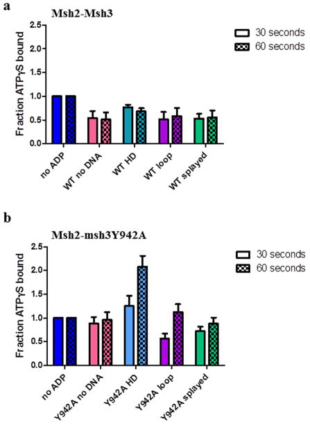
Figure 7. ADP alters ATPγS binding by Msh2-Msh3 and Msh2-msh3Y942A.
Filter binding assays were performed to assess ATPγS binding to (a) Msh2-Msh3 or (b) Msh2-msh3Y942A pre-bound to ADP in the absence of DNA (pink), in the presence of homoduplex DNA (light blue), in the presence of MMR DNA (+8 loop) substrate (purple) or in the presence of 3′NHTR (splayed) substrates (green) after 30 seconds or 60 seconds. The 30 second (left; solid) and 60 second (right; checkerboard) time points are shown side-by-side. The amount of ATPγS bound in the presence of ADP was normalized to the equivalent condition in the absence of ADP (dark blue) and expressed as a fraction. The mean of at least three independent experiments is plotted (± S.E.M.) for each condition.

