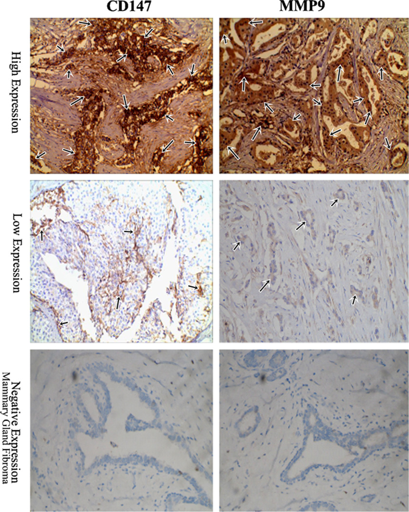
Fig. 1.
Immunohistochemistry (×200) for CD147 and MMP-9 in representative specimens. Positive stain of CD147 and MMP-9 was seen in all 127 cases of tumor samples. High expression of CD147 (n = 61) and MMP-9 (n = 68) was observed, respectively. The predominant pattern of CD147 and MMP-9 staining was cytoplasmic and the cell membrane. All mammary gland fibroma tissue (n = 30) was negative expression for CD147 and MMP-9

