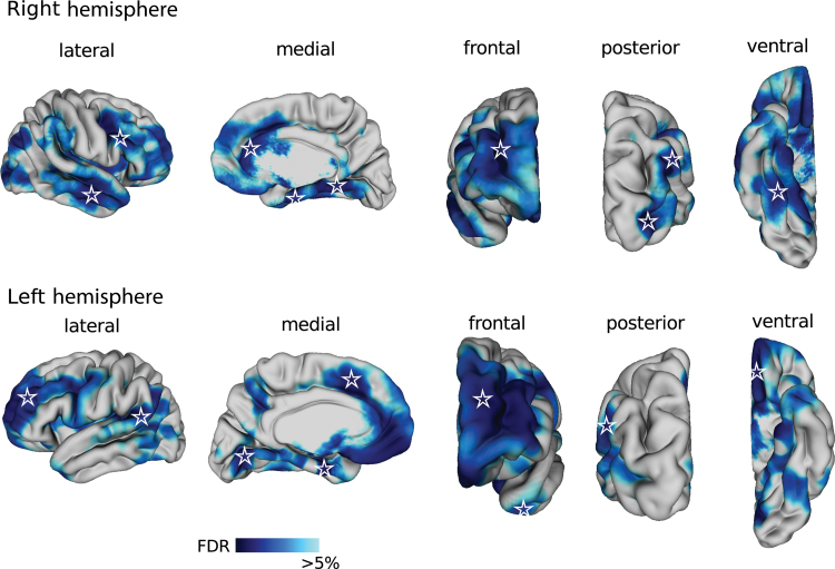
Fig. 1.
Cortical thinning in patients with schizophrenia. Blue color map shows cortical regions that were thinner in patients with schizophrenia after correcting for multiple comparisons at a 5% false discovery rate. Cortical surface of the brain is displayed from the lateral, medial, frontal, posterior, and ventral views in the right (top) and left (bottom) hemispheres. Stars indicate vertices that were used as seeds in the cortical thickness correlation analysis.

