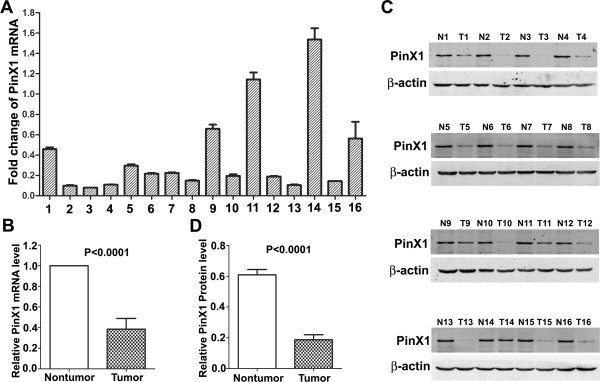
Figure 1.

qRT-PCR and Western blot analysis of PinX1 expression in paired PCa and adjacent normal prostate tissues. (A) Fold changes (2-△△Ct values) by qRT-PCR showed a reduced expression of PinX1 mRNA in the majority of PCa cases (14/16),when compared with paired normal prostate tissues. Expression levels were normalized for GAPDH. (B) Significant differences of PinX1 mRNA expression between the PCa and adjacent normal prostate tissues (P < 0.0001). (C) Western blotting indicated down-regulation of PinX1 protein in PCa tissues (15/16) in comparison with the adjacent normal prostate tissues. β-actin was used as internal control. T, PCa; N, normal. (D) Significant differences of PinX1 protein expression between the PCa and adjacent normal prostate tissues (P < 0.0001).
