
Figure 2.
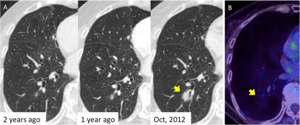
Radiological findings of the nodule. (A) Chest computed tomography (CT) shows an irregularly shaped nodule in the right lower lobe. A tiny nodule at the same site had been found two years earlier. The nodule had since shown a clear tendency to increase in size. The arrow points to the lesion. (B) Positron emission tomography (PET)-CT shows slight accumulation of fluorodeoxyglucose in the nodule (SUVmax = 1.5). The lesion is indicated by the arrow.