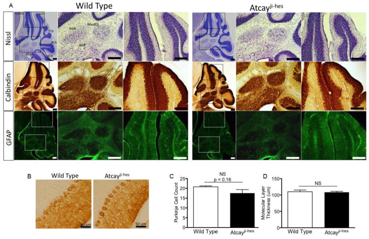
Figure 2. Dystonia in the Atcayji-hes mice develops in the absence of changes in cerebellar architecture or neurodegeneration.
A. Top row. Nissl staining of brain sections from wild-type and Atcayji-hes mice show no evidence of neurodevelopmental abnormalities. Middle row: Calbindin staining for Purkinje neurons shows intact Purkinje neurons with normal molecular layer thickness. Purkinje neuron terminals in the DCN are also intact (middle panel in each row for each genotype). Bottom row: GFAP staining reveals no evidence for gliosis in the cerebellar cortex or DCN suggesting an absence of neurodegeneration. N=3 mice of each genotype. Scale bars = 250 μM. B. Higher magnification images of calbindin staining the Purkinje cell layer shows no evidence of Purkinje cell loss or reduction in dendritic arborization. The lack of changes in Purkinje neuron morphology is summarized in C. and D. Data in the graphs are presented as mean ± SEM.

