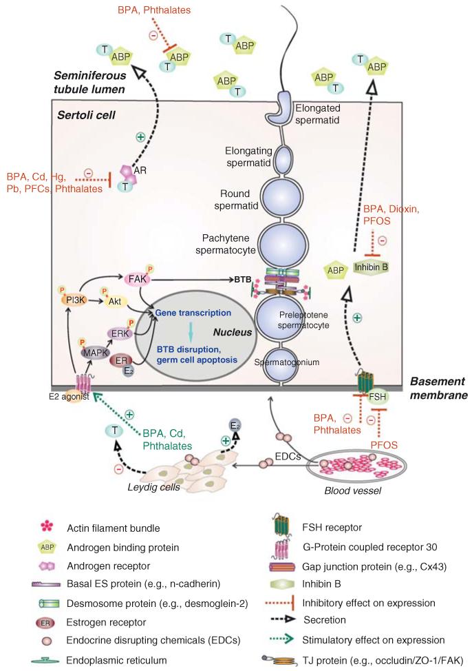
Figure 3. Effects of EDCs-induced changes in the mammalian seminiferous epithelium that disrupts spermatogenesis.
In the mammalian seminiferous epithelium, Sertoli cells are the nurse cells that provide structural and nourishment supports to developing germ cells. The BTB is formed by two adjacent Sertoli cells near the basement membrane. Co-existing tight junction (TJ), gap junction (GJ), and basal ES, which together with desmosome create the BTB. The BTB physically divides the seminiferous epithelium into the basal and the adluminal compartments. Preleptotene spermatocytes differentiate from type B spermatogonia must traverse the BTB to enter the adluminal compartment to differentiate into zygotene and pachytene spermatocytes to undergo meiosis I/II. Ligand binding of FSH onto its receptor as well as androgen onto its receptor stimulate Sertoli cells to secrete androgen binding protein (ABP), which is being used to maintain a high level of testosterone in the tubule lumen to assist sperm maturation. In addition to ABP, Sertoli cells also secrete inhibin B which maintain Sertoli cell normal functioning and increase sperm count/motility. Sertoli cells also secrete a wide range of autocrine, paracrine, and biological factors (such as anti-viral cytokines interferon γ). Sertoli and germ cells also secrete estrogen, which is crucial to maintain male reproductive function via the PI3K/FAK or PI3K/Akt and MAPK/ERK signaling pathways, regulating gene transcription, germ cell apoptosis and BTB function. EDCs have been shown to exert their effects at multiple levels in the seminiferous epithelium depicted herein to affect male reproductive function.

