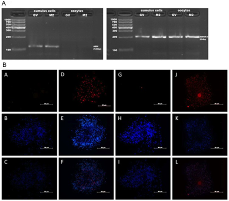
Figure 1. Localization of AMH and AMHR-II by PCR and immunofluorescence staining.
(A) RNA was isolated from oocytes and cumulus cells from 30 COCs that were at GV stage or at MII stage after IVM, respectively. One-microliter amounts of cDNA were used as templates for PCR. (B) COCs after 16–18 h of culture in vitro were stained with mouse isotype IgGs as negative controls of AMH (A) and AMHR-II (G), AMH (D), AMHR-II (J) respectively, followed by TRITC conjugated secondary antibodies and DAPI (B,E,H,K), and merged images (C,F,I,L). Bars = 50 µm.

