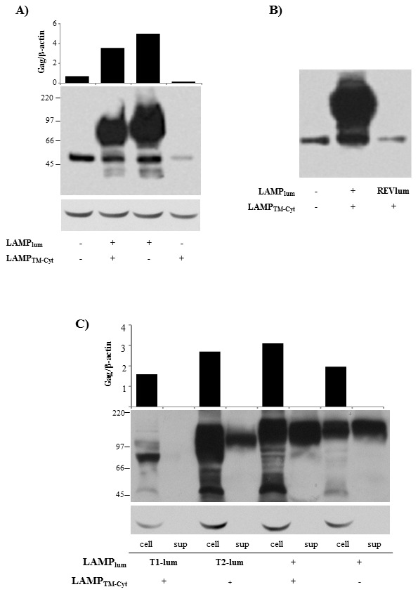
Figure 3. LAMP-mediated increased Gag expression is dependent on LAMP luminal domain.

A–B) HEK293 cells were transfected with the plasmids represented in Figure 2. After 48 h, the amount of Gag protein was analyzed by western blotting, by staining with mouse anti-Gag antibody, followed by HRP-conjugated anti-mouse IgG. The membranes were also probed with β-actin, as a loading control. Bars indicate the ration between Gag expression and b-actin, as measured with ImageJ software. C) HEK293 cells were transfected with the indicated plasmids and, 48 h later, the amount of Gag protein in the cell lysates (cell) and culture supernatants (sup) were analyzed as in (A). Data is representative of four independent experiments.
