
Figure 5. Visualization of the cell free layer in the cremaster muscle by THG with mirror-enhanced signals.
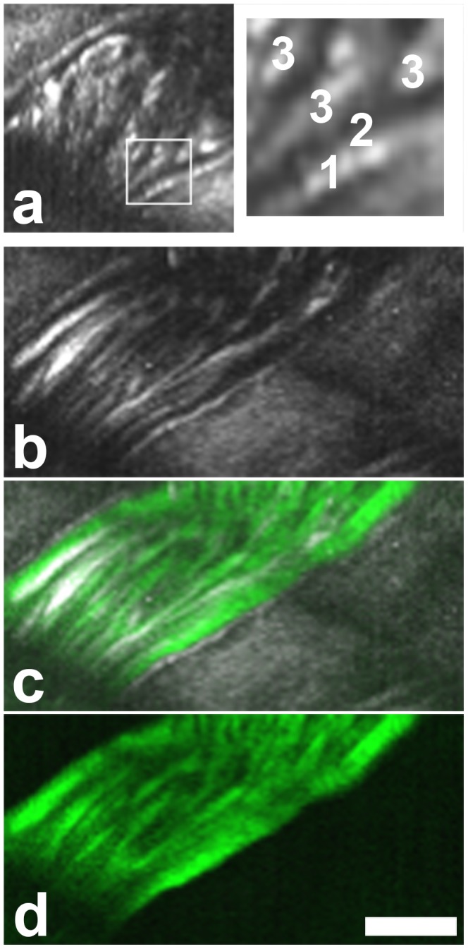
[29] . (a) RBCs are excluded from a gap near the vessel wall. Boxed area magnified on the right. 1, vessel wall. 2, THG free gap. 3, RBC tracks. (b–d) Image of the same area after injection of fluorescently labeled 40 kDa dextran. THG in gray (b,c), dextran fluorescence in green (c,d). The THG free gap is unchanged in width. Contrary to the erythrocytes, this small dextran infiltrates the cell free layer. Scale bar 20 µm. A movie of this vessel is shown in Movie S1.