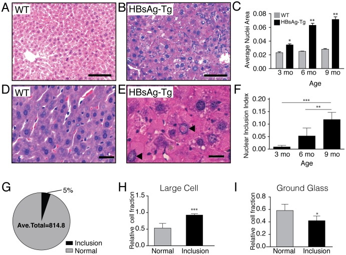
Figure 1. Nucleus size and inclusions in wild type (WT) and nine month old HBsAg-Tg mice.
(A) Hematoxylin and eosin staining of wild type liver, showing hepatocyte with normal nuclei. In contrast, (B) eosinophilic rich nuclear inclusions with larger nuclei can be seen in liver tissue of HBsAg-Tg mice. (C) The size of hepatocyte nuclei increases at 3 months (n = 3), 6 months (n = 3), and 9 months (n = 4) of age compared to wild type controls (n = 2) at each time point. Higher magnification in (D) WT and (E) HBsAg-Tg demonstrates inclusions with eosinophilic material. (F) The ratio of cells with nuclear inclusions compared to normal nuclei increases at 3 months (n = 3), 6 months (n = 3), and 9 months (n = 5) of age. (G) The fraction of hepatocytes with nuclear inclusions was 5% of the total hepatocyte population in HBsAg-Tg mice (n = 5). (H) Large cell dysplasia correlated strongly with hepatocytes having nuclear inclusions (n = 5). (I) Correlation of ground glass hepatocytes with and without nuclear inclusions (n = 5). *,p<0.05; **,p<0.01; ***,p<0.001; ****, p<0.0001, A, B scale bar 100 µm; D, E scale bar 20 µm.

