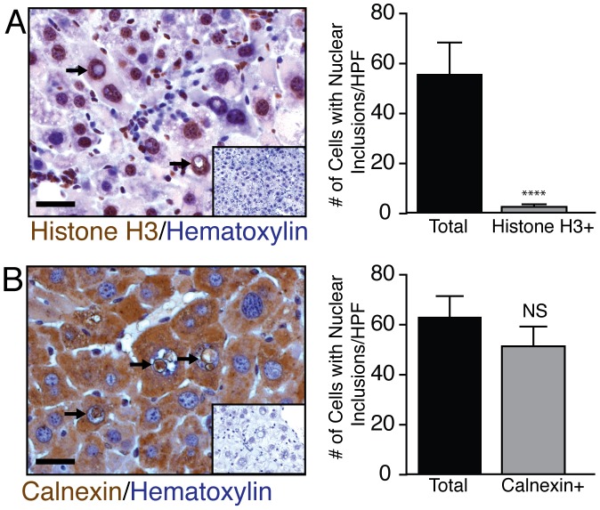
Figure 4. Nuclear inclusions are enriched with endoplasmic reticulum material.
The origin of material within these inclusions was examined by Histone H3 and Calnexin staining. (A) None of the nuclear inclusions expressed histone H3 (n = 5). (B) However, almost all inclusions were clearly positive for calnexin staining (n = 5). Negative controls for Histone H3 and calnexin (no primary antibody) are shown in the insets. scale bar 20 µm.

