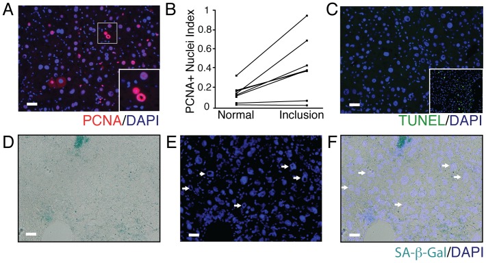
Figure 5. Nuclear inclusions are associated with the proliferative response.
(A) PCNA positive vacuolated nuclei along with positive PCNA “normal” nuclei could be observed in HBsAg-Tg mice. The inset shows a magnified image of PCNA expressing cells with and with out nuclear inclusions. (B) The fraction of PCNA positive with nuclear inclusions was increased in the subset of hepatocytes bearing nuclear inclusions in nine-month-old HBsAg-Tg mice. (C) Cell death as determined by TUNEL assay was not a feature of hepatocytes with nuclear inclusions. The inset shows the carbon tetrachloride induced liver injury model (48 hour post intoxication time point) as a positive TUNEL control. (D-F) Senescence was rarely observed in the HBsAg-Tg at 9 months and did not correlate with hepatocytes containing nuclear inclusions. scale bar 20 µm.

