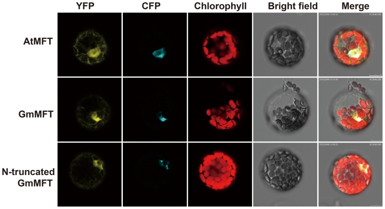
Figure 7. Subcellular localization of GmMFT and N-truncated GmMFT.
YFP represents yellow fluorescent signals of GmMFT:YFP, N-truncated GmMFT:YFP or AtMFT:YFP; CFP represents cyan fluorescent signals of the nuclear protein marker AHL22; Chlorophyll represents chloroplast auto-fluorescence; Bright field represents images from white light; Merge represents merge images of the former four images.

