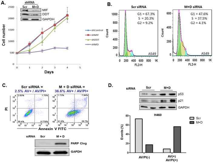
Figure 2. MIF and D-DT depletion results in defects in cellular proliferation, growth, and survival.
A: A549 cells were infected with lentiviral Scr, MIF, D-DT or a combination of MIF + D-DT shRNA. After 72 h, an equivalent number of selected cells were plated in quadruplicate and enumerated for 4 days following plating. B,C: A549 cells were transfected with siRNA oligos as indicated for 96 h. Cell cycle distribution was assessed using FACS analysis of propidium iodide (PI) stained cells (B). Apoptosis was evaluated using FACS analysis of Annexin-V/PI-stained cells and immunodetection of cleaved PARP in lysates (C). D: H460 cells were transfected with siRNA oligos for 72 h followed by immunoblotting of lysates (top panel) or 96 h followed by FACs analysis of Annexin-V/PI-stained cells as in (B). Data shown are representative of 2 (A) or 3 (B,C) independent experiments. ***, p<0.001 by t-test analysis is indicated for individual group comparisons to Scr control.

