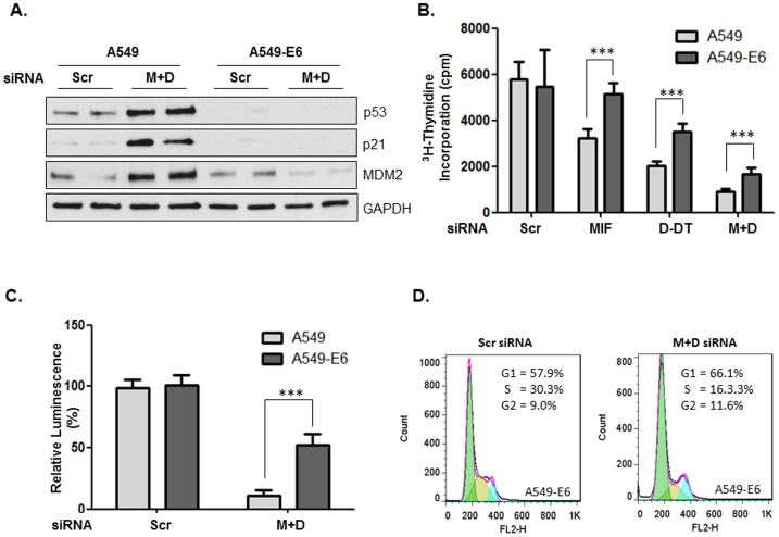
Figure 3. Phenotypic effects of MIF/D-DT depletion are only nominally dependent on p53.
A: A549 and A549-E6 cells were transfected with siRNA oligos as indicated for 72h and lysates were analyzed by immunoblotting. B,C: MIF and/or D-DT were silenced by siRNA transfection as indicated in A549 or A549-E6 cells for 48 h, followed by re-plating into wells of a 96-well plate. Cell proliferation was assessed by a 3H-thymidine incorporation assay (B) and viability was assessed using the Cell-Titer Glo Assay (C). D: MIF and/or D-DT were silenced by siRNA transfection as indicated in A549-E6 cells for 96 h followed by FACS analysis of propidium iodide (PI) stained cells. Data shown are representative of 3 independent experiments. ***, p<0.001 by one-way ANOVA analysis is indicated for individual group comparisons.

