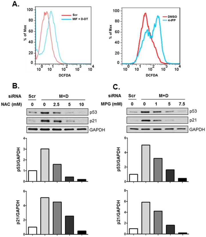
Figure 6. MIF and D-DT regulate p53 in a redox-dependent manner.
A: A549 cells were transfected with siRNA oligos for 72 h or treated with 50 µM 4-IPP overnight as indicated. Intracellular ROS levels were assessed by flow cytometry upon incubation with the fluorescent ROS detector, DCF-DA. B,C: A549 cells were transfected with siRNA oligos as indicated. 48 h later, increasing concentrations of NAC (B) or MPG (C) were added to the cells for an additional 16 h. Lysates were analyzed by immunoblotting. Bio-Rad Quantity One software was used for densitometry and p53/GAPDH or p21/GAPDH densitometry values are depicted in the graph. Data shown are representative of four independent experiments.

