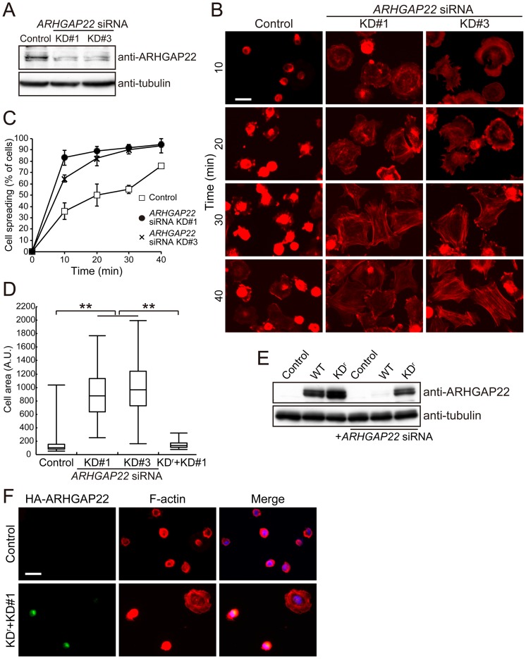
Figure 8. Depletion of ARHGAP22 stimulates cell spreading on fibronectin.
(A) Immunoblot showing that ARHGAP22 is depleted after 48 h of siRNA treatment of C2C12 cells. ARHGAP22 and tubulin were detected by immunoblot using anti-ARHGAP22 and anti-tubulin antibodies, respectively. (B) C2C12 cells were treated with control or ARHGAP22 siRNAs for 48 h and serum-starved. The cells were trypsinized and then plated on fibronectin-coated coverslips and fixed at 10, 20, 30, and 40 min after plating. The cells were stained with phalloidin for F-actin. Scale bar, 20 µm. (C) The percentage of spread cells (n = 200) were calculated and plotted as the mean ± s.e.m. (N = 3). (D) The surface area of spreading cells (n = 100) 10 min after plating was calculated and shown as box and whisker plots. **, p<0.01. Statistical significance was determined by Welch's t-test. (E) HEK cells were treated with control or ARHGAP22 siRNA for 24 h followed by transfection with HA-tagged ARHGAP22 constructs. The cells were cultured for another 24 h. ARHGAP22 and tubulin were analyzed by immunoblot using anti-HA and anti-tubulin antibodies, respectively. (F) C2C12 cells were treated with control or ARHGAP22 siRNA KD#1 for 24 h followed by a transfection with rescue constructs (KDr). The cells were cultured for another 24 h and serum-starved. The cells were fixed and stained with anti-HA antibody for HA-KDr (green) and phalloidin (red). Merged fluorescent images are shown. The cells were also stained with hoechst 33258 for nuclei (blue). Scale bar, 20 µm.

