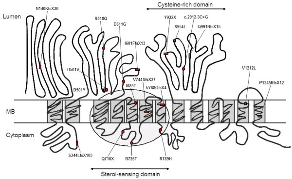
Figure 3.

Localization of mutation identified in 12 Chinese NP-C patients draw on the schematic NPC1 protein model[29]. Red circle: novel exonic point mutations; Red rectangle: novel small insertion/deletions; Green square: reported exonic point mutations; Green rectangle: reported small insertion/deletions.
