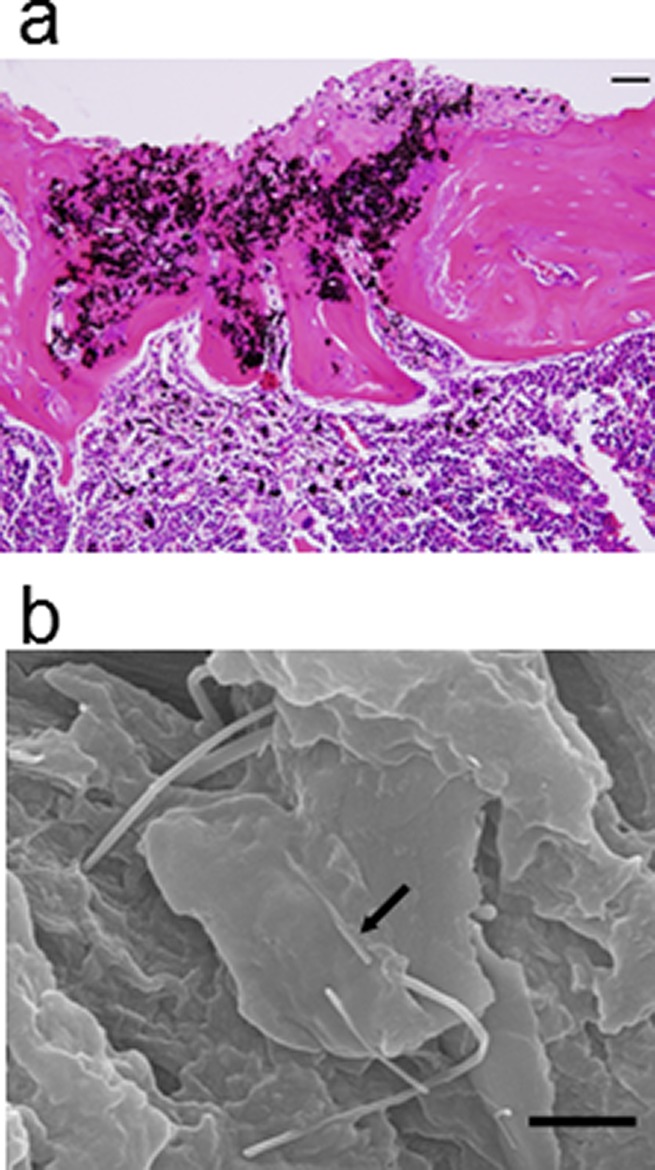
Figure 5.

MWCNTs exhibiting good bone compatibility as they are absorbed in repaired bone without interfering with bone repair. (a) A histological image of a tibia extirpated 4 weeks after surgery for implant of MWCNTs in a pit drilled in tibial diaphysis after incising the anterior surface of a mouse leg. Cortical bone and a medullary cavity were normally formed to the extent of complete bone repair. The MWCNTs were found to have been absorbed in the newly formed bone tissue and enclosed in bone substrate. Hematoxylin-eosin staining. Scale bar = 100 mm. (b) An electron microscopic image of MWCNTs absorbed in repaired bone tissue at 4 weeks. The MWCNTs were found to be in direct contact with bone substrate hydroxyapatite. Scale bar = 1 mm. Reprinted with permission from ref (213). Copyright 2008 John Wiley & Sons, Inc.
