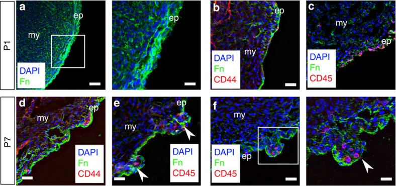
Figure 2. Tertiary epicardial structure forms during early post-natal stages.
At post-natal day 1 (P1), there is no evidence of cell clusters or higher-order structure in the epicardium; Fn staining is diffused throughout (a), and the CD44+ and CD45+ cells that are present do not occupy discrete locations within the epicardium (b,c). By P7, there is evidence of Fn surrounding the forming clusters containing CD44+ (d) and CD45+ cells (e,f; clusters highlighted by white arrowheads). White inset boxes in the left panels are highlighted at higher magnification in neighbouring right panels. ep, epicardium; my, myocardium. Scale bars,: 50 μm (a, left panel); 20 μm (a, right panel); 30 μm (b–f, left panel); 20 μm (f, right panel).

