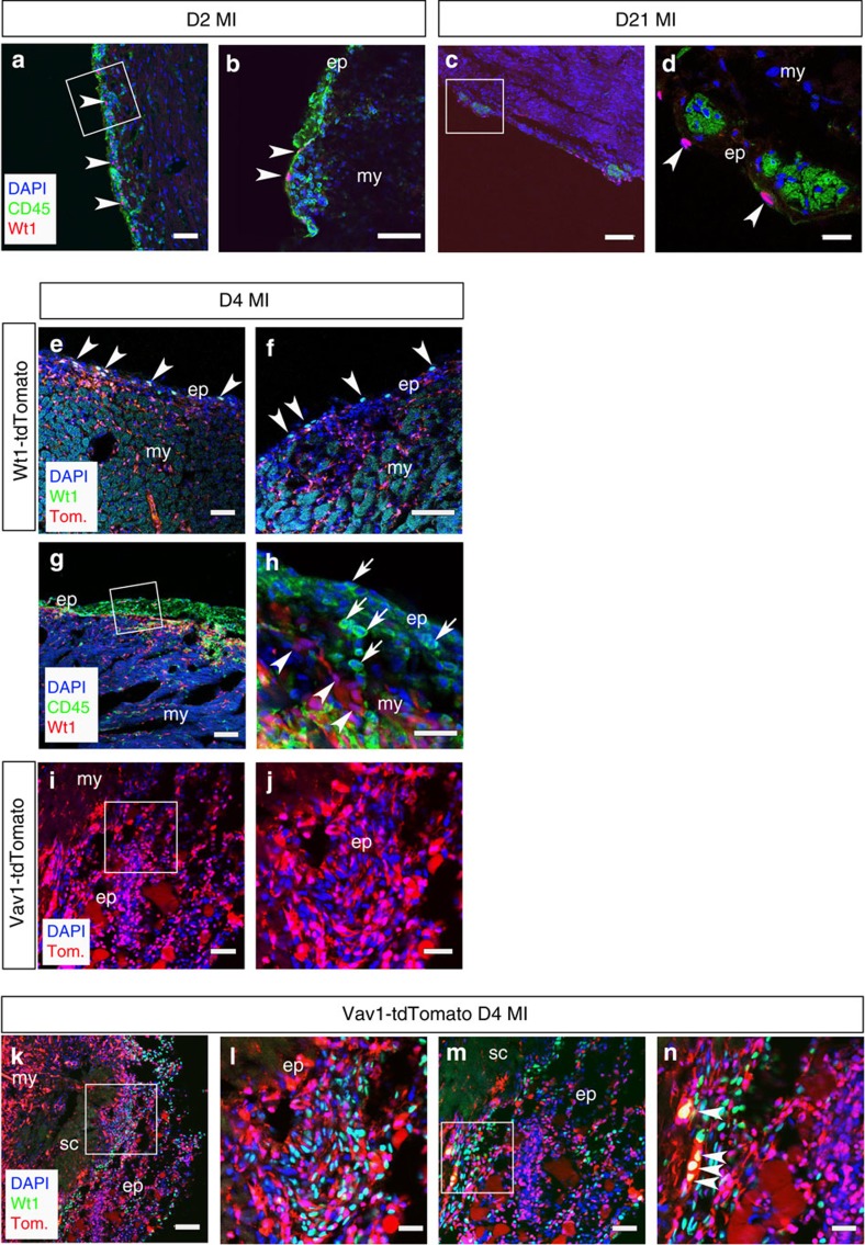
Figure 4. Injury-activated Wt1+ epicardial cells are mutually exclusive from CD45/Vav1+ HCs in the expanded epicardium.
At 2 days post MI (D2 MI), cells re-expressing Wt1 (highlighted by white arrowheads) are distinct from the CD45+ population residing in the expanding epicardium (a,b; white inset box in a, shown at higher magnification in b). At day 21 post MI (D21 MI), Wt1 cells reside proximal to the niche-like clusters (c,d; white inset box in c, shown at higher magnification in d). Wt1CreERT2;R26R-tdTomato labelling and Wt1-antibody staining revealed Wt1+/tdTomato+ cells residing in the expanded epicardium and tdTomato-labelled derivatives within the underlying myocardium (e,f; arrowheads indicate double-positive Wt1+/tdTomato+ cells) relative to an abundance of infiltrating CD45+ cells (g,h; white inset box in g shown in higher magnification in h; arrows in h indicate CD45+ cells and arrowheads indicate Wt1+ cells) and HCs labelled in a Vav1-Cre; R26R-tdTomato reporter line (i,j; white inset box in i, shown at higher magnification in j). At day 4 post MI (D4 MI), Wt1 and tdTomato are expressed in distinct cell populations with a few notable exceptions highlighted by white arrowheads (k–n; white inset boxes in k and m are highlighted at higher magnification in l and n, respectively). ep, epicardium; my, myocardium; sc, scar. Scale bars, 50 μm, (a,c,e,g,i); 100 μm (k,m); 20 μm (b,d); 30 μm (f,h,j,l,n).

