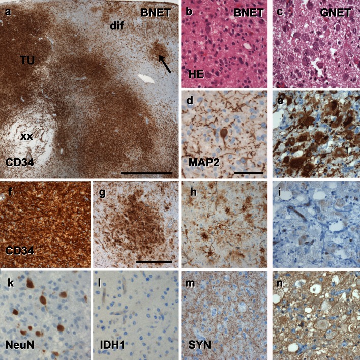
Fig. 4.
The spectrum of histomorphological and immunohistochemical hallmarks in LEATs with a glio-neuronal phenotype (A–B–C terminology). a BNET, immunoreactive for CD34 class II epitope (mAB QBend10, hematoxylin counterstaining; first three columns). Three different patterns can be distinguished. Pattern 1: (TU)—the bulk tumor is densely stained for CD34 (magnification in f). Pattern 2: clusters of tumor cells are visible in adjacent cortical areas (arrow, magnification in g). Pattern 3: diffusely infiltrating CD34-positive cells can be found in distant remote areas from the bulk tumor (magnification in h). Note, that patterns 2 and 3 may be interpreted as FCD ILAE Type IIIb when not using CD34 immunohistochemistry. b, c Routine histology stainings (H&E) reveal a biphasic pattern in BNET and a predominant gangliocytic patter in GNET. d, e Immunohistochemistry for the embryonic MAP2 epitope (MAP2e) is helpful to identify the neuronal component. Compared to diffusely infiltrating gliomas, the glial component does not label for Map2e (see Figs. 5, 6). i no CD34 immunoreactivity is visible in GNETs. k–n Supplementary markers are helpful to distinguish LEAT entities. NeuN labels dysplastic neurons in BNETs (k). IDH1 staining is always recommended as it is detectable only in neoplastically transformed glial cells (diffuse astrocytomas AII and oligodendroglioma OII; see Figs. 5, 6), but not in BNET (l). Synaptophysin (SYN) may be helpful to visualize the neuronal component in BNET (m) and GNET (n). Scale bar in a 2 mm, d 50 µm applies also to b, c, e, i–n. Scale bar in g 200 µm, applies also to f–h

