Fig. 9.
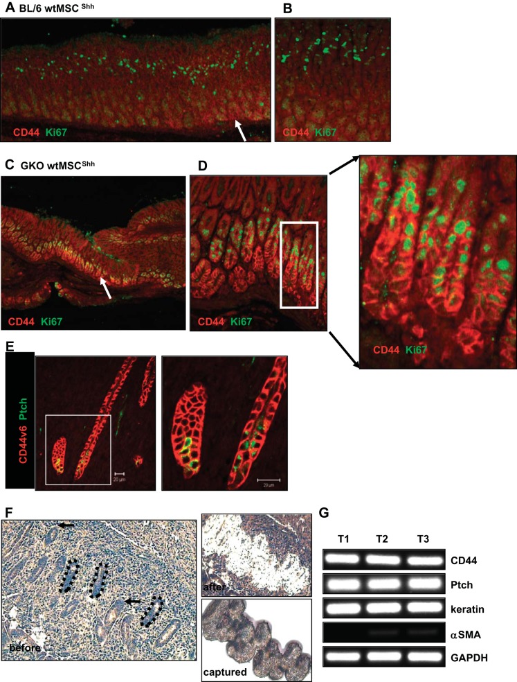
Expression of CD44 and Ki67 in stomachs collected from BL/6 and GKO recipient mice. Representative images of immunostaining using antibodies specific for CD44 (red) and Ki67 (green) using stomach sections collected from BL/6 recipient mice (A) transplanted with wtMSCShh cells (higher magnification in B) and GKO recipient mice (C) transplanted with wtMSCShh cells (higher magnification in D). Arrows show CD44-positive cells. E: immunostaining of CD44v6 (red) and Ptch (green) in GKO recipient mice transplanted with stMSCvect cells. F: paraffin-embedded sections collected from gland-like epithelium within the tumor stroma were stained and the ultraviolet cutting laser was used to first ablate the cells surrounding the region (before and after). An infrared laser was then used to capture the cells from surface pit, neck, and base epithelium (captured). G: gene expression of CD44, Ptch, cytokeratin 20 (keratin), and α-smooth muscle actin (α-SMA) was analyzed by qRT-PCR using RNA collected by Laser Capture Microdissection from 3 representative tumors (T1, T2, and T3) in GKO recipient mice transplanted with stMSCvect cells.

