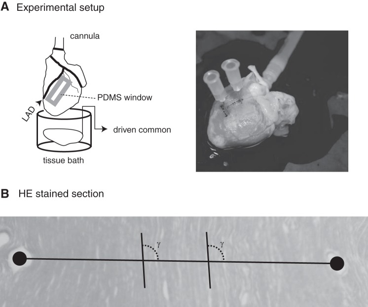
Fig. 2.
A: schematic diagram of experimental setup (left) and photograph of 1 heart into which fiducials were placed at the end of an experiment. B: cropped hematoxylin and eosin (HE)-stained section with line between centroids of fiducials used to identify γ for orientation of electrodes relative to myocyte axes. PDMS, polydimethylsiloxane.

