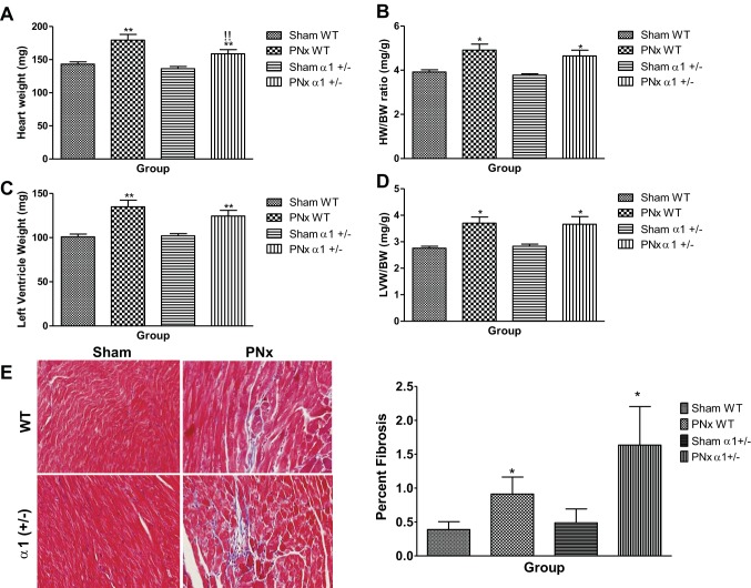
Fig. 2.
PNx induces cardiac hypertrophy and fibrosis in WT and α1+/− mice. Heart weight (HW) and left ventricle weight (LVW) (A and C) were measured after mice were euthanized, and the heart weight/body weight ratio (HW/BW, C) and LVW/BW ratio (D) were calculated based on the final body weight before euthanasia. Both HW/BW and LVW/BW data indicate a significant increase in cardiac hypertrophy. E: Masson's trichrome staining in left ventricle tissue from experimental mice: representative images from each group taken by an Olympus FSX100 microscope with a ×20 lens (left) and quantification data (n = 8 in each group; right). *P < 0.05, significantly different than sham of same genotype; **P < 0.01, significantly different than sham of same genotype; !!P < 0.01, significantly different than PNx of WT.

