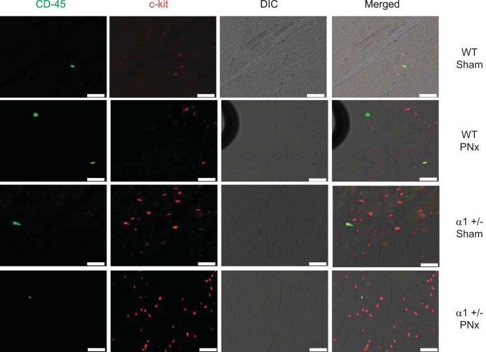
Fig. 8.
Coimmunostaining of CD45 and c-kit in heart tissue. Left ventricle tissues fixed in 4% formaldehyde solution were used for immunofluorescent staining with anti-c-kit antibody and anti-CD45 antibody as described in materials and methods. Data show that over 90% of c-kit-positive cells are CD45 negative, indicating that most of the c-kit cells are not of hematopoietic origin. Scale bar is 25 μm.

