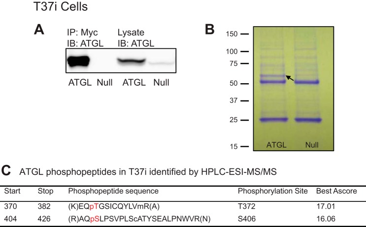
Fig. 3.
Identification of ATGL phosphorylation sites in T37i brown adipocytes. T37i adipocytes were infected with either Ad-Myc-ATGL or Ad-null virus. A: Myc-ATGL was detected in cell extracts and anti-Myc immunoprecipitates by immunoblotting. B: immunoprecipitated proteins were resolved by SDS-PAGE and stained with Coomassie blue dye. The band for Myc-ATGL protein is indicated by the arrow. C: results from HPLC-ESI-MS/MS analysis. The preceding p indicates phosphorylated amino acid. Sites with Ascore of >13 have a P value of <0.05 and >95% certainty for correct phosphorylation site localization. Sites with Ascore of >19 have a P value of <0.01 and >99% certainty for correct phosphorylation site localization (3, 11, 28).

