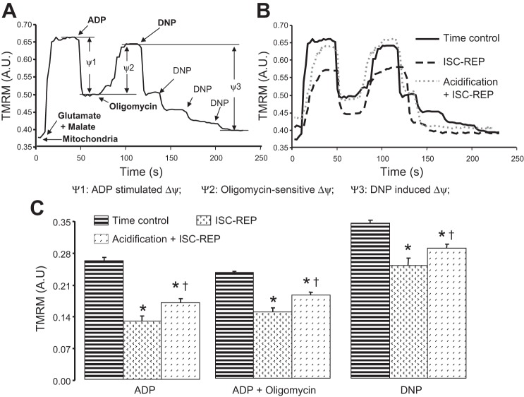
Fig. 6.
Acidic reperfusion preserved mitochondrial inner membrane integrity assessed by the membrane potential indicator TMRM measured following more prolonged periods of reperfusion. A: a representative trace of TMRM fluorescence as an indicator of mitochondrial inner membrane potential. The addition of ADP stimulated oxidative phosphorylation with a depolarization of inner membrane potential. Membrane potential was restored when oligomycin was used to inhibit oxidative phosphorylation at complex V. Dinitrophenol (DNP), an uncoupler, resulted in a depolarized inner membrane potential. B: representative traces of TMRM fluorescence from mitochondria isolated from time control, untreated ischemia-reperfusion, and acidic reperfusion-treated hearts. The changes of fluorescence intensity of TMRM in the presence of ADP, oligomycin, and DNP were compared in C. Ischemia-reperfusion (ISC-REP) significantly decreased the relative inner membrane potential in the presence of ADP, oligomycin, and DNP compared with the time control group. Acidic reperfusion treatment substantially improved the relative measurement of inner membrane potential as a functional index of mitochondrial integrity compared with mitochondria from untreated hearts. Values are means ± SE; *P < 0.05 vs. time control; †P < 0.05 vs. untreated; n = 5 in each group.

