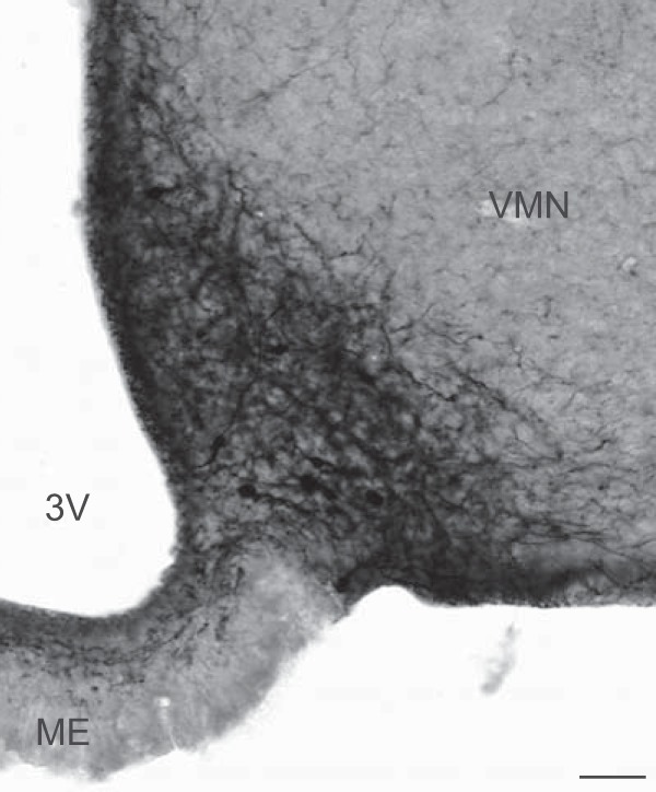
Figure 3.

Photomicrograph of EGFP immunocytochemistry in the arcuate nucleus of a Tac2-EGFP female mouse. Sections were visualized with nickel-intensified DAB. Intensely labeled cell bodies were obscured by numerous beaded axons and dendrites. Axons extended to the median eminence, periventricular zone, ependymal layer, and the pial surface. The adjacent ventromedial nucleus was devoid of EGFP-ir somata. See Supplemental Figures 1 and 2 for photomicrographs of EGFP immunohistochemistry of other brain regions. 3V, third ventricle; ME, median eminence; VMN, ventromedial nucleus. Scale bar, 50 μm.
