
Figure 4.
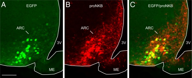
Representative photomicrographs of EGFP-fluorescence (A, green) and pro-NKB-immunofluorescence (B, red) in the arcuate nucleus of an OVX, Tac2-EGFP mouse. Nearly 80% of EGFP neurons in the arcuate nucleus were labeled by the pro-NKB antibody (C, yellow). ARC, arcuate nucleus; ME, median eminence; 3V, third ventricle. Scale bar in A, 100 μm (applies to A–C).