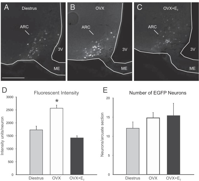
Figure 5.
EGFP fluorescence in the arcuate nucleus of Tac2-EGFP mice. Fluorescent arcuate neurons in the OVX mice (B) appeared qualitatively brighter than neurons in diestrous (A) and OVX + E2 (C) mice. D, Semiquantitative image analysis revealed a significant increase in the average fluorescent intensity units per neuron in the arcuate nucleus of OVX mice. E, The mean number of fluorescent neurons counted in a unilateral arcuate nucleus section was not significantly different between groups. 3V, third ventricle; ARC, arcuate nucleus, ME, median eminence. Values are mean ± SEM, n = 5–6 mice/group. *, significantly different from diestrous and OVX + E2 mice (one-way ANOVA with Tukey's post hoc test).

