
Figure 1.
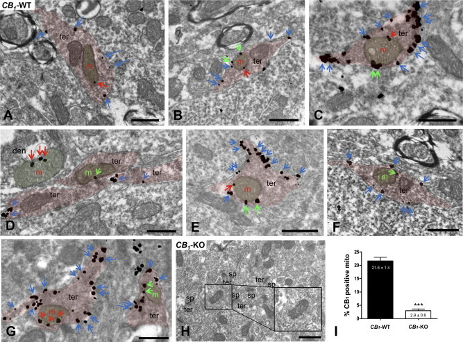
Immunoelectron detection of CB1 receptors in neuronal CA1 hippocampal mitochondria by a goat anti-CB1 C-ter31 antibody combined with a pre-embedding silver-intensified immunogold method. In CB1-WT (A–G), CB1 immunoparticles are on presynaptic terminal (ter) membranes (blue arrows) and on mitochondrial (m) membrane segments close to (distance ≤80 nm; green arrows) or far away from (distance ≥80 nm; red arrows) other membranes. Note in (D), red arrows pointing to silver metals on a dendritic (den) mitochondria. (H) The CB1 pattern is abolished in CB1-KO. Scale bars: 0.5 μm. sp: dendritic spines. (I) Semi-quantitative analyses of the proportion of CB1 immunolabeled mitochondria in CA1 hippocampi from CB1-WT and CB1-KO mice. Mitochondria with particles distant from other membranes (≥80 nm) were only considered. Data are expressed as mean ± S.E.M. ***p < 0.001 as compared to WT.