
Figure 2.
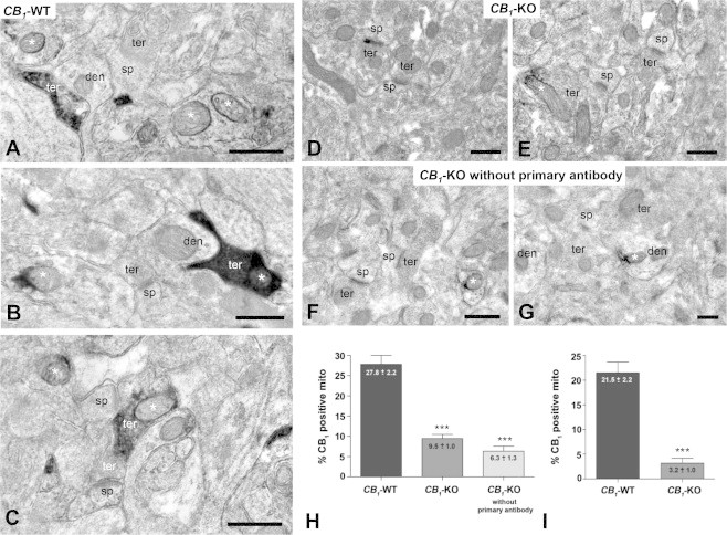
Ultrastructural localization of CB1 receptors in mitochondrial sections with the immunoperoxidase DAB–Ni method using a goat anti-CB1 C-ter31 antibody. (A–C) In CB1-WT mouse, CB1 immunoreactivity was observed in mitochondrial sections (asterisks), some of them were contained in CB1-positive synaptic terminals (black immunoreaction product). (D and E) Absence of CB1 immunodeposits in mitochondrial sections and synaptic terminals of CB1-KO mouse. Only some scattered mitochondrial sections display DAB–Ni precipitates (asterisk in E). (F and G) Unspecific immunodeposits on mitochondrial sections (asterisks) and dendritic profiles of CB1-KO mouse when the primary antibody was omitted. (H) Semi-quantitative analyses of the proportion of CB1 immunopositive mitochondrial sections in CB1-WT and CB1-KO mice, and in CB1-KO mice without primary antibody. (I) Semi-quantitative analyses of the proportion of CB1 immunopositive mitochondrial section in CB1-WT and CB1-KO mice after subtraction of the percentage value obtained in CB1-KO mice without primary antibody. Data are expressed as mean ± S.E.M. ***p < 0.0001 according to chi-square test. ter: presynaptic terminals; den: dendrites; sp: dendritic spines. Scale bars: 0.5 μm.