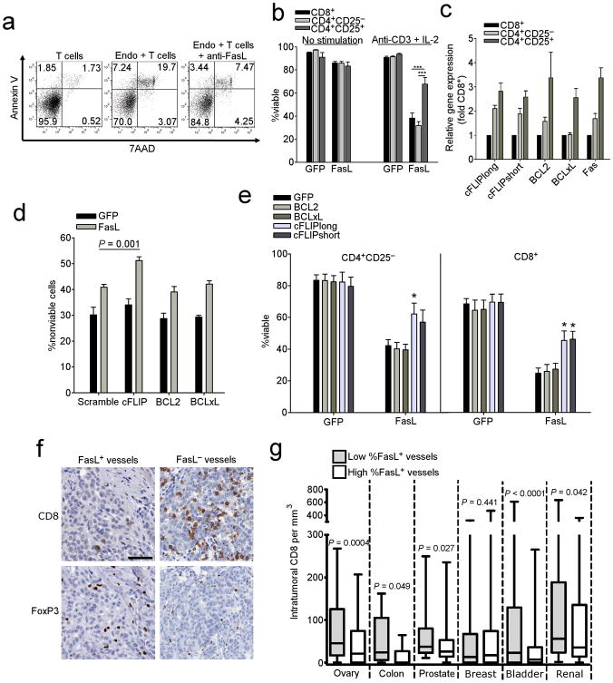
Figure 2. Endothelial FasL kills T cells.
(a) Viability of T cells following co-culture with endothelial cells isolated from human ovarian tumors incubated at a 3:1 ratio with or without anti-FasL antibody. Gated on CD3+ T cells. (b) Killing of normal donor T cell subsets by HMEC-1 cells transduced with either GFP or FasL. Data are means ±SEM pooled from three independent experiments using unique healthy donors. ***P < 0.001, values determined by Student's t test. (c) Expression of anti-apoptotic genes in human T cell subsets as determined by qRT-PCR. Data are means ±SEM pooled from six independent experiments using unique healthy donors. (d) Killing of shRNA transduced Tregs by HMEC-1 cells transduced with either GFP or FasL. Data are means ±SEM pooled from four independent experiments using unique healthy donors. P values determined by Student's t test. (e) Killing of T cells transduced with anti-apoptotic genes by HMEC-1 cells transduced with either GFP or FasL. Data are means ±SEM pooled from four independent experiments using unique healthy donors. P values determined by Student's t test. (f) Representative images of CD8 and Foxp3 cells in sections taken from subjects with ovarian cancer expressing either FasL+ or FasL− vessels. Original magnification, 400×; scale bar, 50 μm. (g) Intratumoral CD8 T cell counts from human TMAs associated with percentage of FasL+ vessels. High/low determinations based on median values. P values determined by Student's t test.

