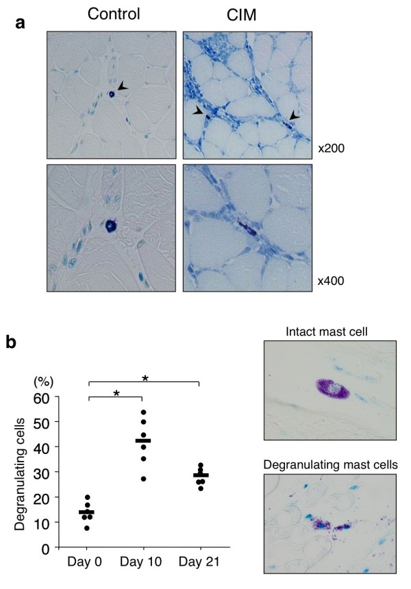
Figure 2.

Histological analysis of skeletal muscle in mice with C protein-induced myositis (CIM). (a) Mice were immunized with murine skeletal C protein fragment or vehicle as described in Materials and methods. Twenty-one days after the immunization, specimens of proximal muscle were stained with toluidine blue. Arrowheads indicate mast cells. (b) Kinetic analysis of mast cell degranulation. Mice were immunized with murine skeletal C protein fragment, and at indicated days after the immunization, the frequency of degranulating mast cells in skeletal muscle was evaluated as described in Materials and methods. Dots show the percentage of degranulating mast cells of individual mice, and horizontal bars show mean percentages of degranulating mast cells. n = 6 mice at each time point, *P <0.01.
