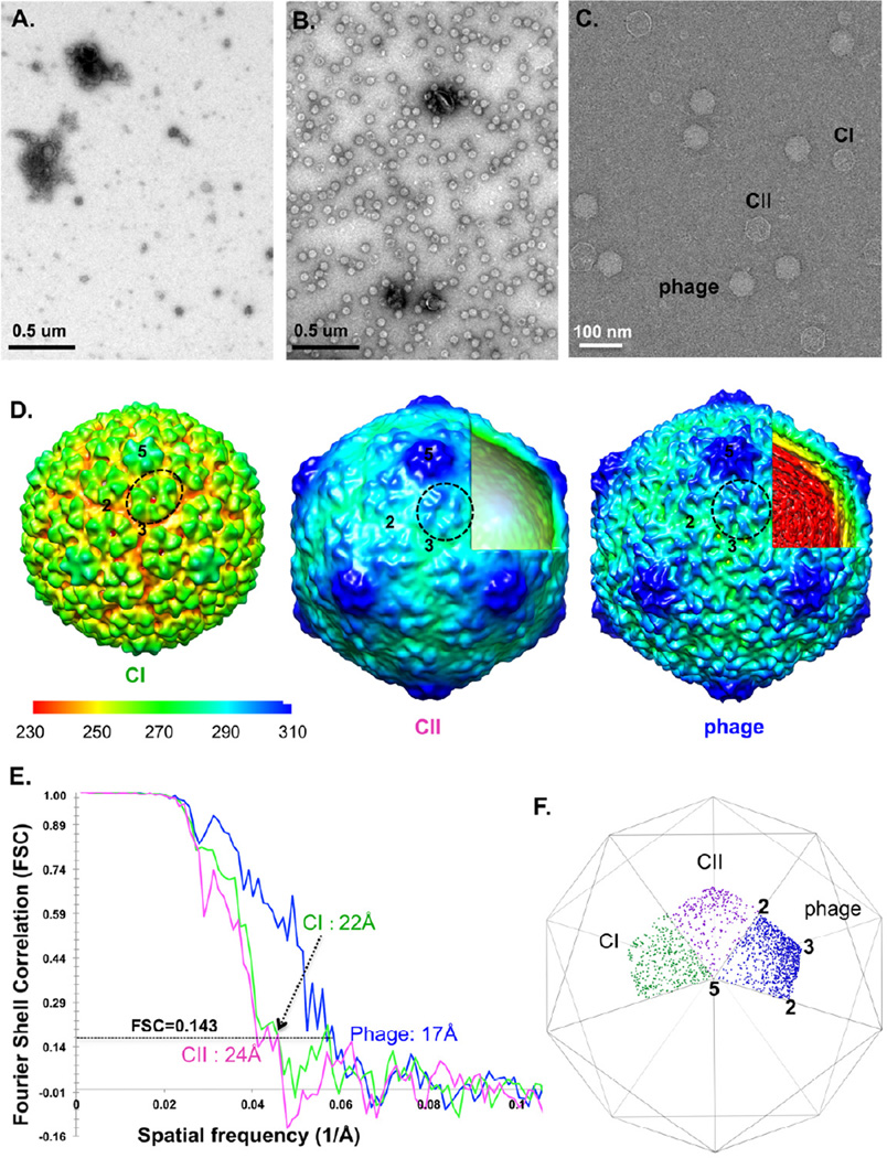
Figure 2. His-T7 bacteriophages directly captured from a non-purified cell lysate by SPIEM grids coated with anti-His antibodies.
Negative staining TEM images of a control grid (A) and anti-His antibodies coated grid (B) show the specific “trapping” and “concentrating” of target particles. (C) cryo-SPIEM images of His-T7 particles directly from a cell lysate. Three states of T7 phages: initial procapsid (CI), the empty capsid II (CII) and the DNA-filled mature phage. Single particle cryo-EM 3D reconstruction of the three states of His-T7 (D) and the estimated resolutions using gold-standard FSC=0.143 criterion (E). The figures: “2”, “3” and “5”, indicate the icosahedral two-, three- and five-fold axes, respectively. One capsid protein hexamer of each map was circled. (F) shows the Euler angle distributions of CI, CII and mature phage particles on cryo-SPIEM grids.

