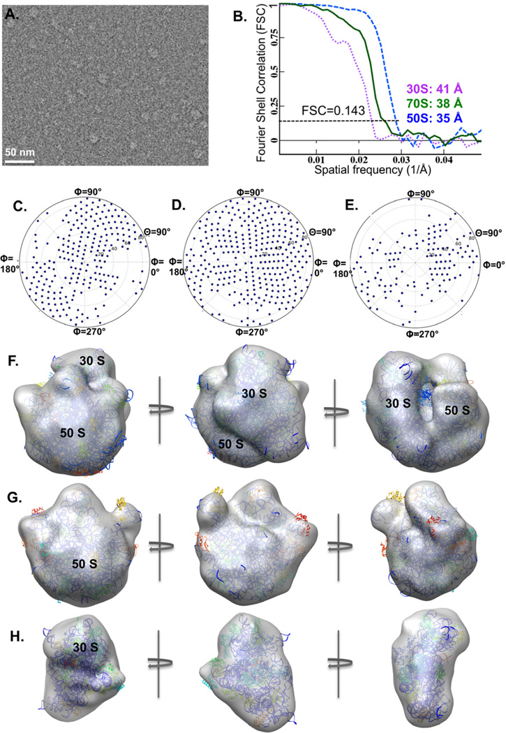
Figure 3. His-tagged E. coli ribosomes directly imaged using the cryo-SPIEM approach with anti-His antibodies.
(A) An anti-His antibody based cryo-SPIEM image of E. coli ribosomes from a crude cell lysate. (B) Fourier shell correlations curves of the 70S, 50S and 30S ribosomes captured on the cryo-SPIEM grid. And the Euler angle distributions of the 70S (C), 50S (D) and 30S (E) are plotted. The 3D reconstructions of the 70S, 50S and 30S ribosomes fitted with corresponding crystallographic structures (PDB-1ml5, 3e1b, 4b3m) are shown in (F), (G) and (H), respectively.

