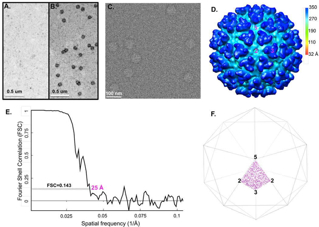
Figure 4. Native enveloped Sindbis virus directly captured from infected BHK-21 cell culture supernatant for single particle cryo-EM 3D reconstruction.
Negative staining images show that Sindbis particles specifically bind to the grid coated with anti-E2 antibodies (B) but not to the grid without anti-E2 (A). (C) shows one representative cryo-SPIEM images of Sindbis particles directly from the 8-fold concentrated cell culture supernatant. (D) Single particle 3D reconstruction of the captured particles. “2”, “3” and “5” represent the icosahedral two-, three- and five-fold axes, respectively. (E) Fourier shell correction of the reconstruction. (F) plots the Euler angles of the Sindbis particles captured on the cryo-SPIEM grid.

