
Figure 2.
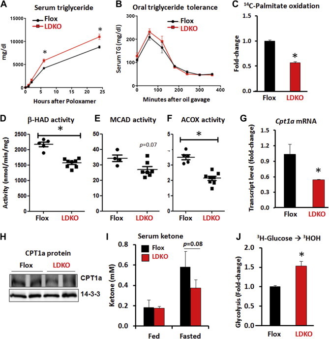
The loss of ACC activity decreases hepatic fat oxidation. (A) Hepatic triglyceride production assessed by serum triglyceride levels at time points after i.p. treatment of 1 g/kg lipoprotein lipase inhibitor, Poloxamer 407 (n = 5 LDKO vs. n = 4 flox). (B) Oral triglyceride tolerance assessed by serum triglyceride levels at various time points after a bolus of safflower oil (n = 5). Data expressed as mean ± SEM; *p < 0.0001, multiple unpaired t-test with correction using Holm-Sidak method. (C) Rate of palmitate oxidation in isolated primary hepatocytes (*p < 0.001, Mann–Whitney, seven independent experiments). Enzyme activity assays of (D) 3-hydroxyacyl-CoA dehydrogenase (β-HAD), (E) media-chain acyl-CoA dehydrogenase (MCAD), and (F) peroxisomal acyl-CoA oxidase (ACOX) in liver tissue (*p < 0.05, Mann–Whitney, n = 7 LDKO vs. n = 5 flox). (G and H) Hepatic CPT1a transcript levels and protein expression.14-3-3 serves as a protein loading control. (I) Serum ketone levels at fed and fasted states (Mann–Whitney; n = 5 flox vs. n = 10 LDKO). (J) Rate of glycolysis in isolated primary hepatocytes (*p < 0.01, Mann–Whitney, five independent experiments). Data expressed as mean ± SEM.