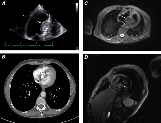
Fig. 1.

A) Transthoracic echocardiogram (apical 4-chamber view) shows a solid mass in the basal intraventricular septum. B) Cardiac computed tomogram shows a heavily calcified mass in the basal septum. Cardiac magnetic resonance images show the mass C) with isodensity similar to that of the myocardium (triple-inversion cardiac sequence) and D) by delayed gadolinium enhancement.
