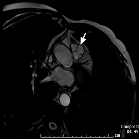
Fig. 1.

Cardiovascular magnetic resonance steady-state free-precession sequence shows closure (arrow) of 3 pulmonary valve cusps of equal size and 1 smaller, poorly formed cusp. Pulmonary valvular motion was also apparent throughout the cardiac cycle.
Supplemental motion image (2.4MB, m1v) is available for Figure 1.
