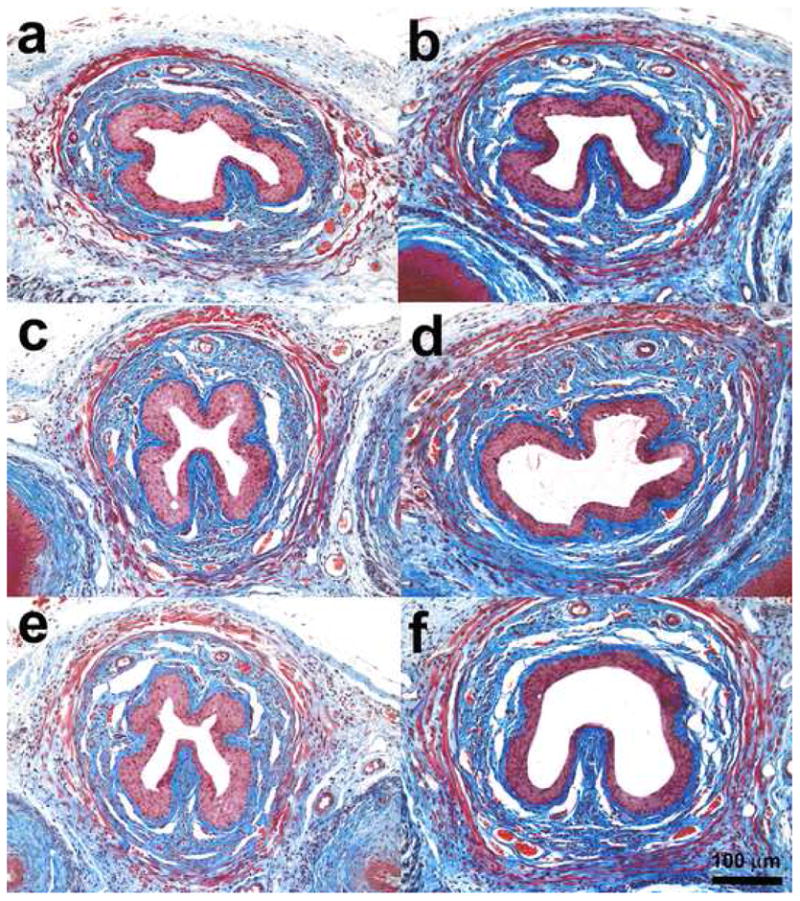
Fig. 2.

Images (Masson's trichrome staining) of transverse sections of the midurethra at (a) 0 d after vaginal distension (VD), (b) 4 d after VD, (c) 10 d after VD, (d) 20 d after VD, (e) 4 d after pudendal nerve transection, and (f) 4 d after sham VD.
