
Figure 4.
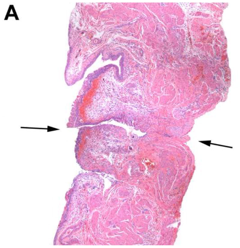
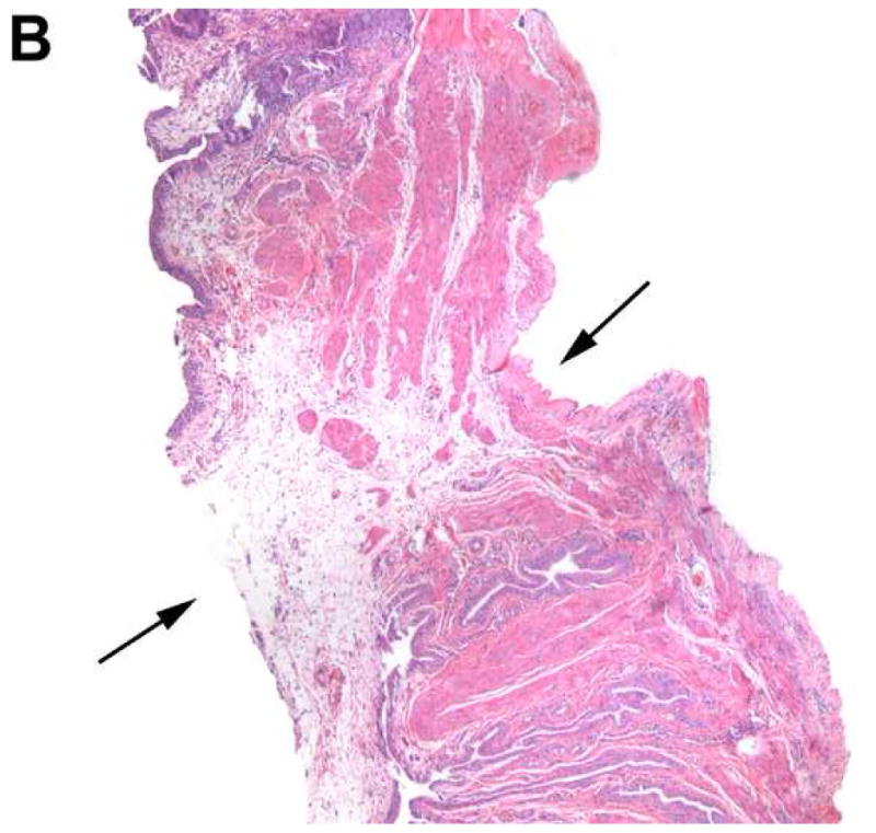
Histology of the bladder in a device-implanted rat. H & E staining of the bladder under light microscopy (40×) illustrates the defect created by the device (black arrow) and mild inflammatory changes in adjacent areas 1 day (A) and 4 days (B) after implantation.