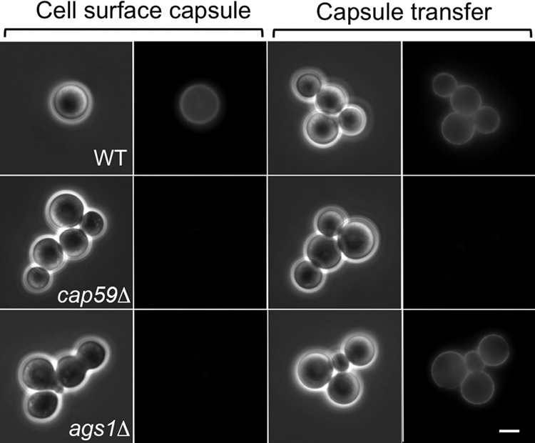
FIG 1.

Detection of cell surface capsule and capsule transfer. (Left two columns) Endogenous surface capsule of the indicated strains was probed by staining with the anti-GXM monoclonal antibody 3C2. (Right two columns) The cap59Δ acapsular strain was used as the acceptor for exogenous capsule from CM of the indicated strains, washed, and stained similarly. The left panel of each pair is a differential interference contrast (DIC) image, and the right panel is an immunofluorescence (IF) image. All images are at the same magnification. Bar = 5 μm.
